Acontia
In C. polypus, one region of the cnidoglandular band of each septal filament is known as the acontia. (Ruppert, 2004) Acontia are nematocyst dense, threadlike extensions of mesenterial filaments that are highly ciliated (Rodríguez et al., 2012, Hessinger and Lenhoff, 1973). Acontia develop at the pedal end of the mesenterial filaments, with the free ends laying in the coelenteron.(Ruppert, 2004) They are usually emitted from the anemone when irritated, exiting through the cinclides located on the column or via the mouth along with coelenteric fluid. The acontia of C. polypus is salmon pink in colour and is made up of most basitrich nematocysts (Salleo et al., 1990).
Specimens were collected from Frenchman’s Beach, Stradbroke Island. Acontia were then collected from the specimens by initiating the retraction-deflation response and cutting them with a scalpel. The acontia were then mounted on a slide to inspect under a microscope. Attempts at sectioning the acontia were unsuccessful, so full samples were used.
|
|
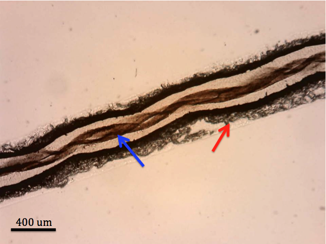
| Figure 1: Mounted acontia with half of the epithelium removed in order to see the coiled mesenterial filament inside. The blue arrow points to the mesenterial filament. The red arrow points to the dense numbers of basitrichs on the surface of the acontia. |
|
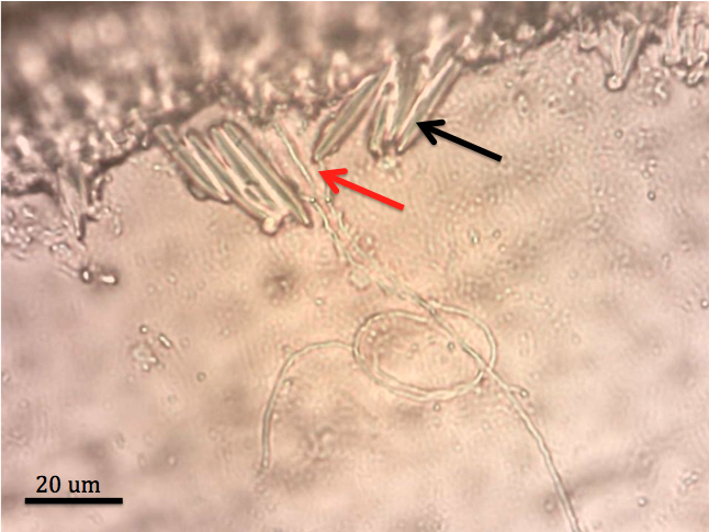
| Figure 2: A closer look at the surface of the acontia. Red arrow points to fired basitrichs, their fired tubules extending away from the acontia. Black arrow points to unfired basitrichs. |
|
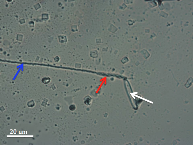
| Figure 3: A fired basitrich. White arrow points to the empty capsule of the basitrich. Red arrow points to the barb dense section of the tubule. Blue arrow points to the extended tubule. |
|
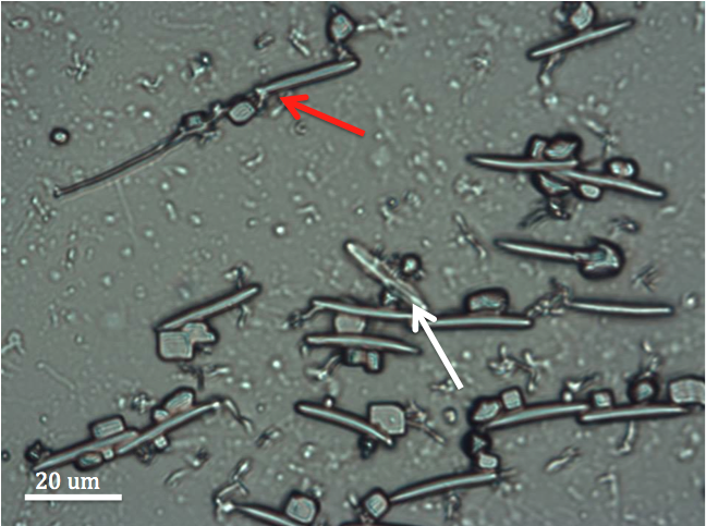
| Figure 4: Fired and unfired basitrichs from acontia. Red arrow points to fired basitrich. White arrow points to unfired basitrich - the tubule is visible inside the capsule of the nematocyst. |
|
|
| Video of C. polypus after irritation, showing the acontia being expressed through the cinclides. |
|